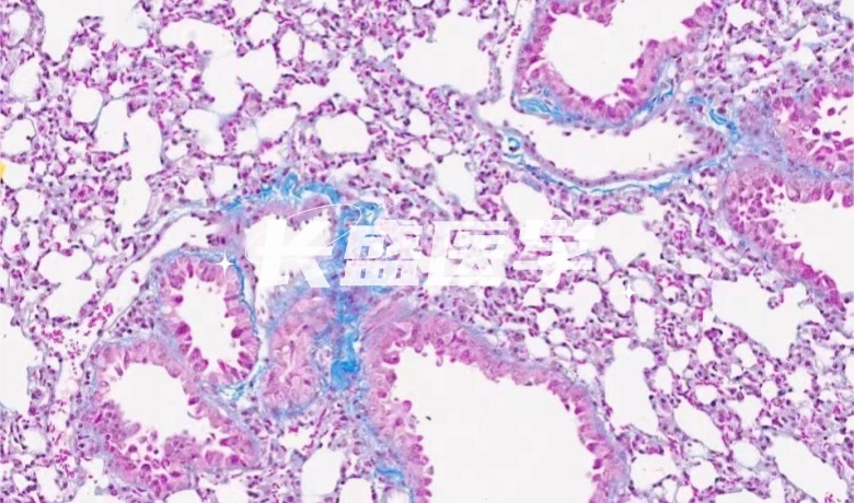
Masson staining
When Masson is dyed, the collagen fibers appear blue or green as they are stained with the dye aniline blue or brilliant green. Conversely, muscle fibers appear red because they are colored by acid fuchsin and rosin dyes. This difference is closely related to the molecular size of the anionic dye and the permeability of the tissue to the dye.
Technical principle
Masson staining is a common histological staining method that stains collagen fibers, muscle fibers, and red blood cells by using anionic dyes of different sizes to address differences in tissue permeability and chemical composition. In this method, collagen fibers are colored blue or green with a larger molecule of anionic dye; muscle fibers are colored red with a medium-sized anionic dye; and red blood cells are colored with the smallest molecule of anionic dye. This staining method has a high clinical application value, because it can quickly and accurately show different tissue components, so as to help medical professionals to diagnose and treat different diseases.
Real Experimental Research Hundreds of Detection Experiments 6 Experimental Platforms
More experiments
Real experimental research data is unique
Plasma experiment service
Cold atmospheric plasma (CAP) is an emerging tumor treatment method that has been shown to be selective for many types of cancer cells, providing new opportunities for effective treatment of various cancers. CAP is a plasma generated at room temperature and atmospheric pressure, and has shown great potential for biomedical applications due to a favorable combination of reactive physical and chemical species, such as UV radiation, electrons, free radicals, ions, and excited molecules. Recently, it has been found that plasma activated medium (PAM) produced by exposure of aqueous media to CAP is as effective as direct CAP in inhibiting cancer cells, thereby increasing the range and flexibility of plasma-based therapy.
Real experimental research data is unique
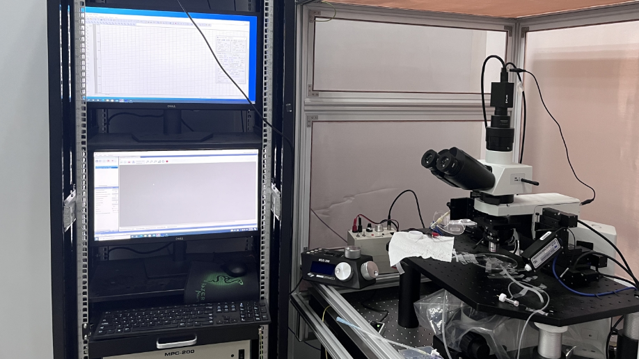
Electrophysiological Experiment Service
Electrophysiological platform: The brain slice patch clamp system consists of amplifier, digital-to-analog converter, micro-operation, electrode drawing instrument, microscope and perfusion system. With a special glass micropipette adsorbed on the cell surface membrane, and then the implementation of the membrane voltage clamp, can measure the current of the magnitude of pA generated by the opening of a single ion channel. It can also be used to detect the cell excitability (action potential and model index), ion channel (potassium sodium calcium ion) and synaptic transmission (excitatory synaptic transmission and inhibitory synaptic transmission) in the brain slice of the desired site of drug/experimental treatment.
Real experimental research data is unique
Single Cell Sequencing
The single-cell sequencing platform provides you with comprehensive single-cell multi-omics services, including whole transcriptome (WTA) analysis, targeted transcriptome (TTA) analysis, full-length analysis of human V(D)J, full-length analysis of mouse V(D)J, and VDJ CDR3 analysis. Our protein group has 500 commercial Abseq antibodies: covering human/mouse immune-related Marker, AbSeq (human) immune detection combination, 30 kinds of personal immune Marker; At the same time, it provides multi-sample detection, including 12 kinds of human multi-sample labels, 12 kinds of mouse multi-sample labels, 6 kinds of nuclear multi-sample labels, genome scATAC-seq detection and CRISPR/Cas9 detection. At present, our single-cell sequencing platform involves many fields such as tumor typing, targeted drug use, immunotherapy, animal and plant embryonic development, and the occurrence and development mechanism of cardiovascular diseases. No matter what field you are engaged in, our services can meet your needs and provide comprehensive support for your research.
Real experimental research data is unique
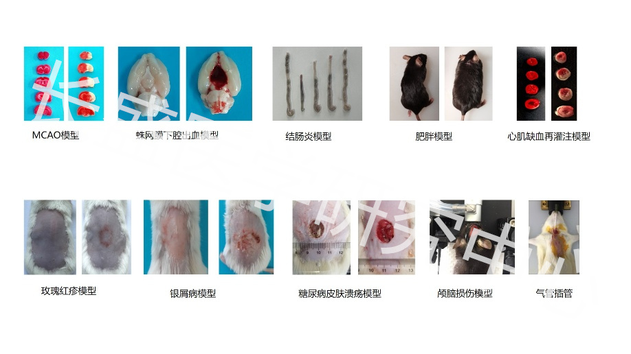
model animal
Changsheng medical animal modeling platform provides outsourcing services for animal disease modeling for customers, and can undertake the establishment of animal models related to various basic medical research, such as tumor, liver disease, respiratory disease, nervous system disease, cardiovascular disease, urinary system disease, endocrine disease, orthopedic disease, immune system disease, blood disease, gynecological disease, skin disease and other animal models. The modeling process supports field trips to ensure the quality and reliability of animal experiments.
Real experimental research data is unique
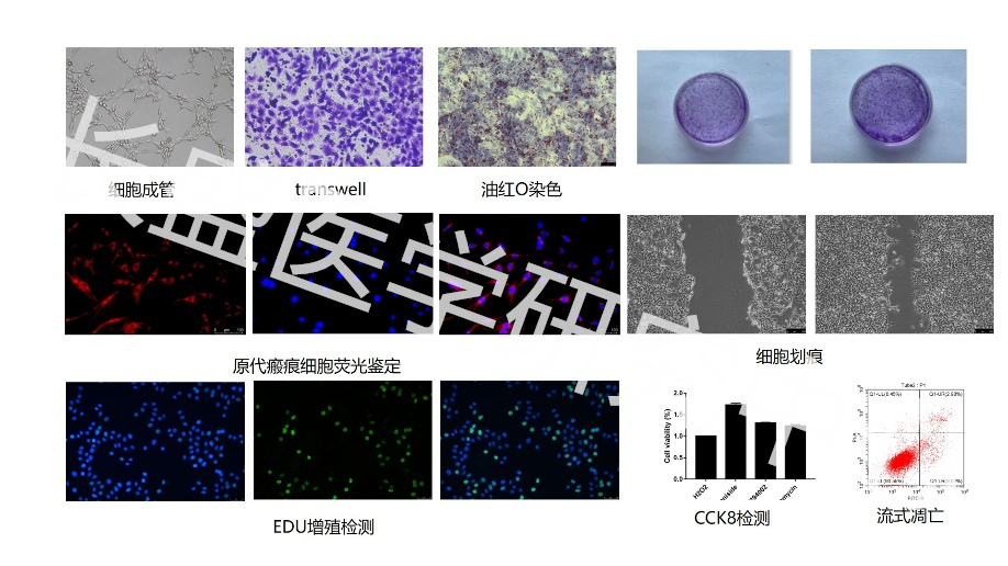
cell experiment
We provide an advanced cell experiment platform, which can detect the proliferation of MTT, CCK8, Edu and other cells, and can detect the invasion and migration ability of cells. We also provide experimental services such as plate cloning, soft agar cloning and comet assay, which can develop drug-resistant strains, construct cell lines and detect the function of tumor cells as needed. In addition, we offer an extensive library of cell resources that you can purchase to make your research results more reliable.
Real experimental research data is unique
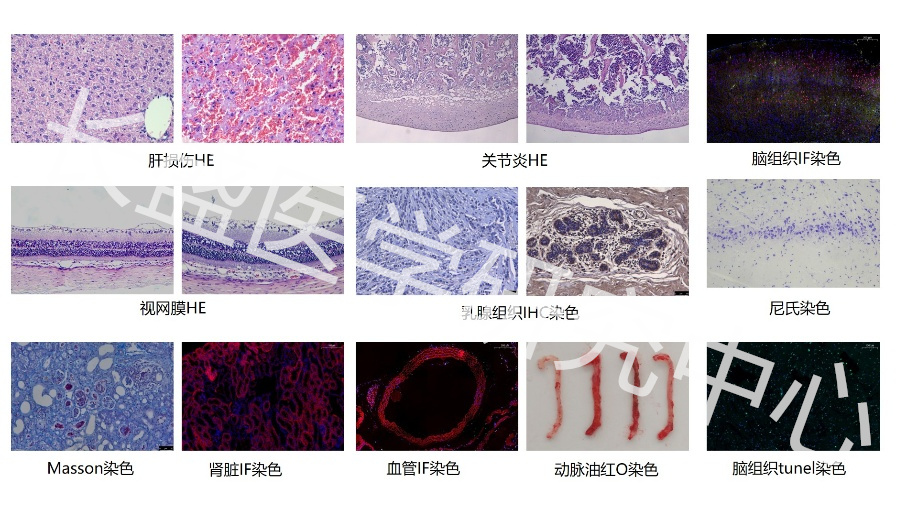
tissue staining
Our pathology platform provides customers with comprehensive biological sample processing services. You can entrust us to carry out paraffin section, section scanning, fluorescence scanning, HE staining, and various special staining items, such as Masson, oil red O, PAS, safranin O, ALP, TRAP, toluidine blue, Tunel staining, methanol Congo red staining, safranin solid green staining, TTC staining, immunohistochemistry/fluorescence, in situ hybridization, etc.
Real experimental research data is unique
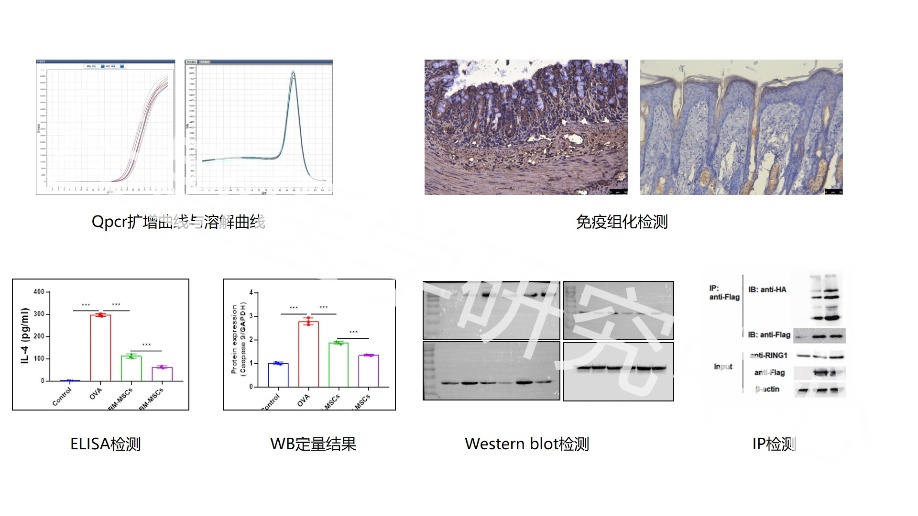
expression detection
We have a first-class expression detection platform, can provide real-time fluorescence quantitative PCR, DNA methylation detection, Western blot, ELISA, SNP and mycoplasma detection and other professional services,. Our testing services cover the whole country, providing the perfect solution for the outsourcing of biomedical experiments. Our commitment is to provide customers with 100 percent true and reliable service, sign relevant legal agreements with customers, and provide permanent after-sales service.